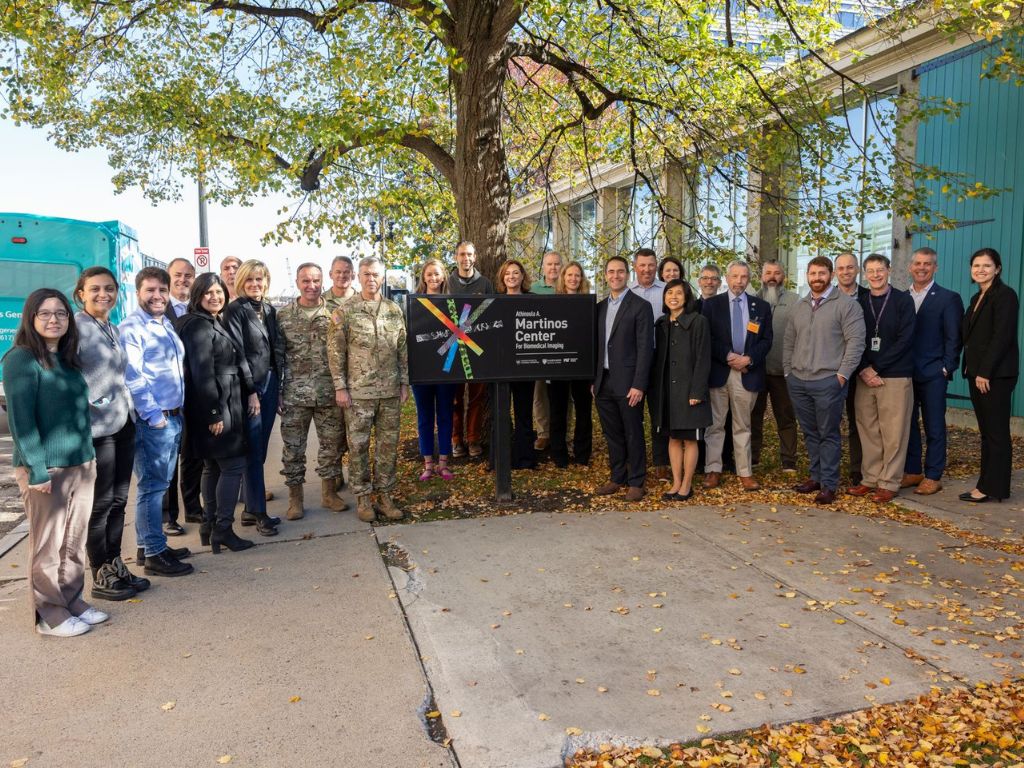
From Innovation to Impact: Disseminating EPTI Across the Neuroimaging Community
The next-generation MRI acquisition technique overcomes major limitations of conventional methods.
The next-generation MRI acquisition technique overcomes major limitations of conventional methods.
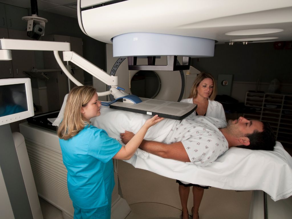
MRI scans taken before prostate cancer treatment may already contain clues about which patients are most at risk for the disease returning after radiation. In a new study, Onofrio A Catalano and colleagues analyzed pretreatment prostate MRIs and… MRI Clues Could Help Predict Prostate Cancer’s Return After Radiation
Doctors often use an invasive catheter inside an artery to get continuous blood pressure readings during surgery, but that can be uncomfortable and risk complications. In a new study, Zahra Einalou, Maria Angela Franceschini and colleagues tested… FlexNIRS Offers Needle-Free Way to Track Blood Pressure
Understanding how different parts of the brain communicate requires precise maps of neural activity. In a new study, Tommi Raij and colleagues improved the way scientists interpret data from magnetoencephalography, or MEG — a technology… A Faster, Clearer Way to See Brain Activity with MEG
PET scans can reveal inflammation in the brain, but they’re costly and require injecting a small amount of radioactive material. In a new study, Marco Loggia and colleagues developed an artificial intelligence system that can create “synthetic”… Generating PET Images — Without the Radiation
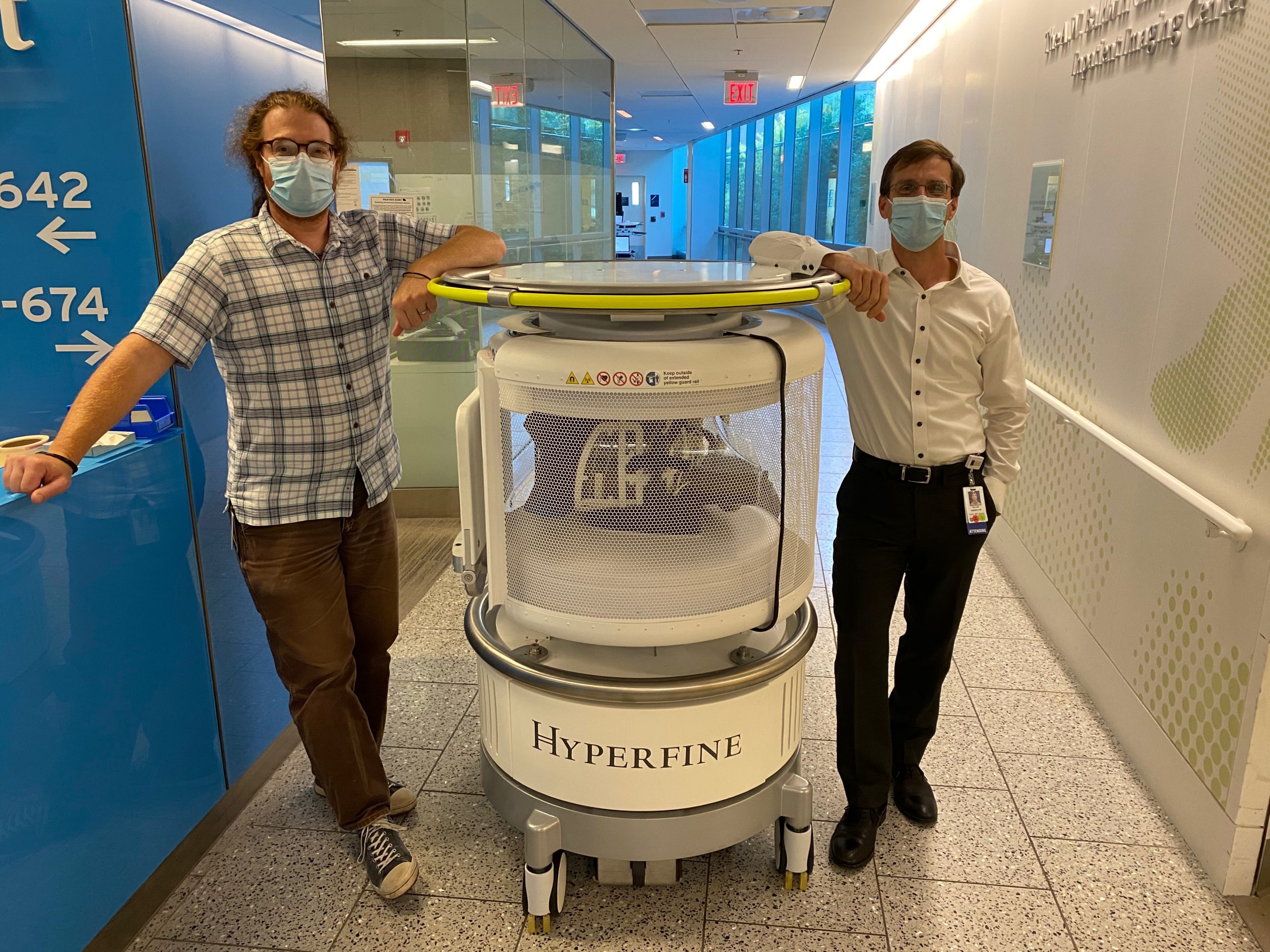
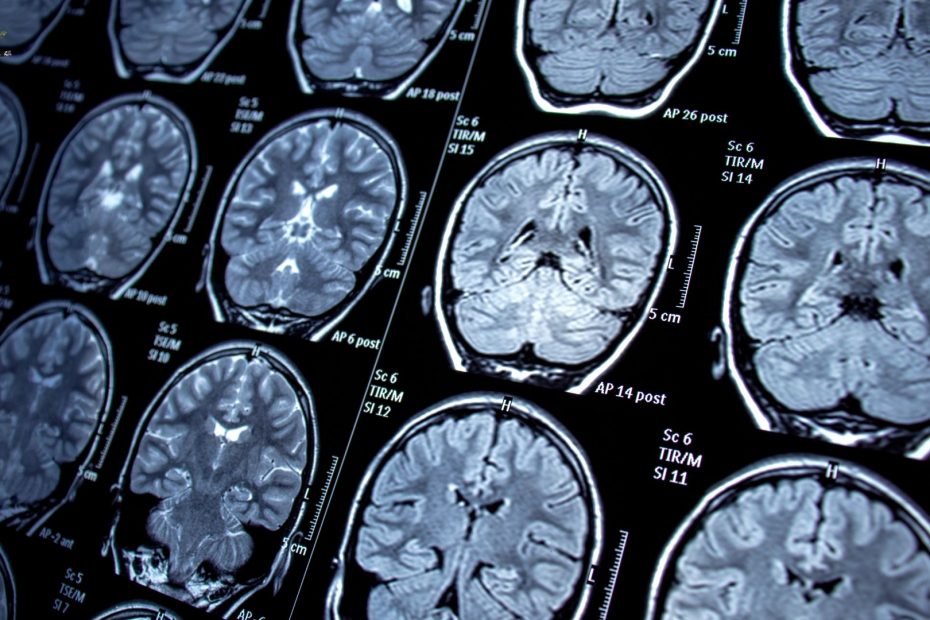
Matthew Rosen was part of an effort to address barriers to the successful implementation of pMRI for brain research.

The award will fund development of a wearable device for women that monitors the brain’s system that clears waste during sleep.

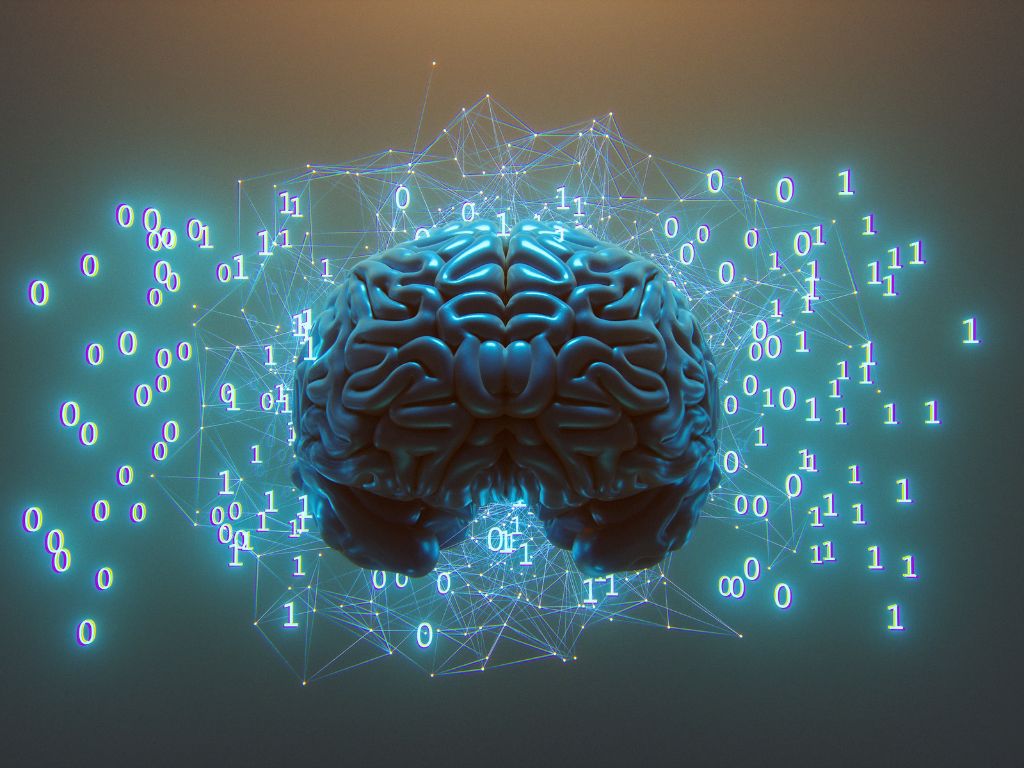
The tool will make it easier for researchers without the technical expertise to take advantage of AI.
Malte Hoffmann, PhD, and colleagues have developed a new, AI-based approach to image registration that is more robust, accurate, and faster than existing methods.

Recent research by Matthew Sacchet, PhD, and colleagues establishes strong proof of concept for studying advanced meditation in controlled neuroimaging settings.

The work was conducted by the Center’s Tal Kenet, PhD, and colleagues.

The trial also demonstrated the capabilities of collagen PET imaging.
In a study of 30 active-duty United States SOF personnel, researchers found that increased blast exposure was associated with structural, functional, and neuroimmune changes to the brain and a decline in health-related quality of life.

Martinos researchers Daphne Holt, MD, PhD, Faye McKenna, PhD, and colleagues have identified a potential marker of loneliness, which could help understand the effects of loneliness on the body and even how to reverse them.
Martinos researchers have developed a method for whole-brain segmentation of longitudinal MRI scans that generalizes to different scanners and MRI sequences, does not put any constraints on the number or timing of follow-up scans, and can segment white matter lesions simultaneously.
The center’s Randy Buckner and colleagues have provided direct support for a third somatomotor map in the vermis of the cerebellum by using intensive, repeated functional MRI scanning of individuals performing movements across multiple body parts.
The Center’s Caterina Mainero and colleagues review the interplay of fibrinogen and coagulation factors with neuroinflammation in multiple sclerosis.
A study by the Center’s Leo Cheng and colleagues suggests that metabolomic changes detected with an advanced nuclear magnetic resonance (NMR) spectroscopy technique can differentiate between patients with benign prostate biopsies who will or will not receive a prostate cancer diagnosis over the following years.

Recent studies from the Department of Radiology hav sought new ways to help reduce the incidence of a broken heart (and to ensure a healthy cardiovascular system).
Jyrki Ahveninen and colleagues believe sparse brain activity, where only a few neurons in a population fire at a given time, might contribute to the fingerprint patterns of activity evident in multi-voxel pattern analysis results from functional MRI — and give rise to perceptual, emotional and cognitive functions.